Low resolution
Technology
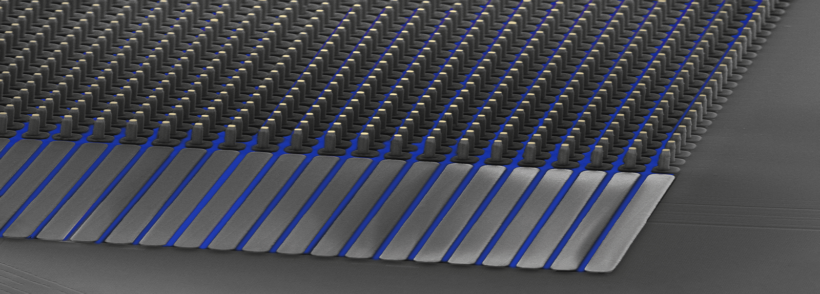
CorePlate™ 3D
A new dimension for Brain-on-chip design
We empower scientists to venture into the unknown
Standard MEA

CorePlate™ 2D
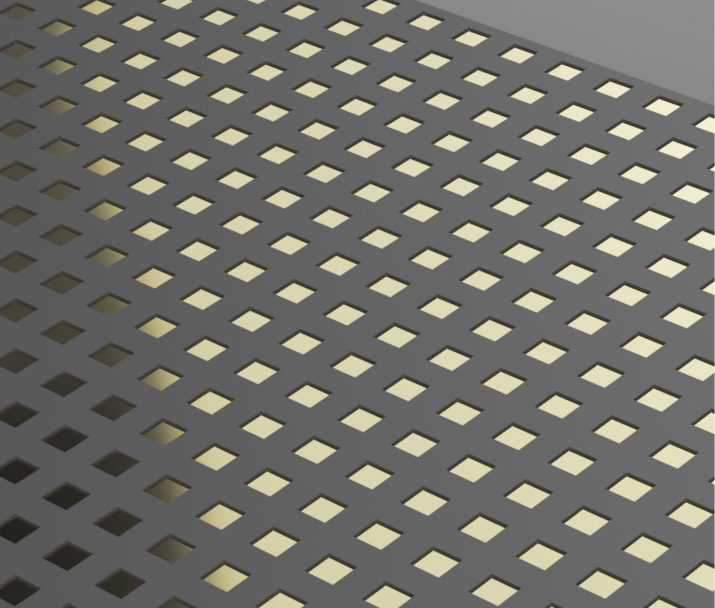
High resolution
CorePlate™ 3D
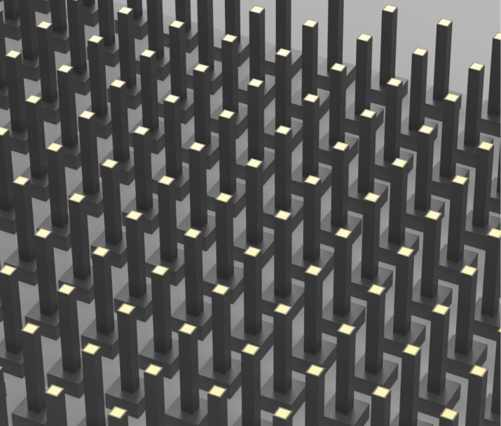
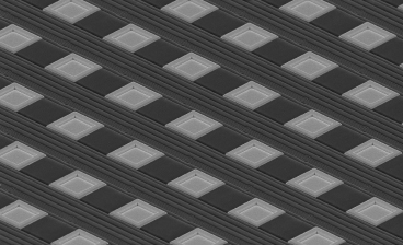
High resolution in 3D
Increased complexity and data depths
3Brain builds technology to face life’s complexity head-on

-
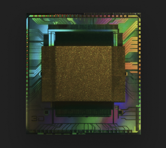
– First 3D CMOS-chip
-
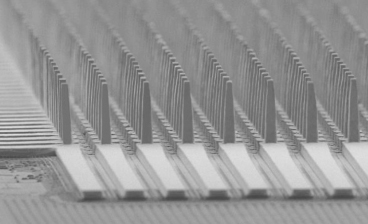
– 4,096 gold-coated sensory electrodes
-
– Bio-compatible polymer isolation
-
– Microfluidic channels
-
– Bidirectional cell-electronic interface
-
– On-chip signal processing
CorePlate™ 3D was developed with and for neuroscientists
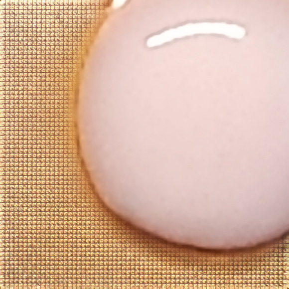
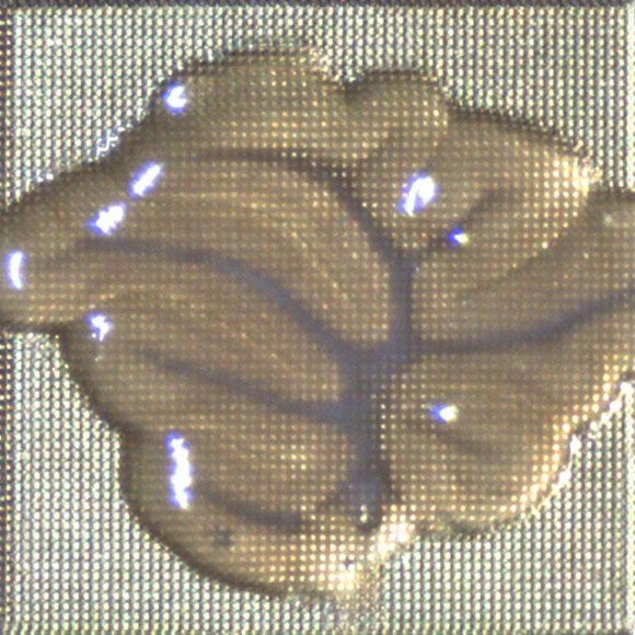
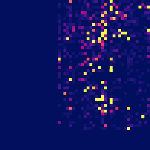

Explore in vitro organoids & ex vivo brain slices
CorePlate™ 1W -3D 38/60/90 enables the investigation of 3D cellular networks at unprecedented depths and resolution. Our real-time biosignal processing microchip broadens the concept of label-free functional imaging beyond optical approaches.
Our bidirectional pixel array technology empowers researchers to communicate with cells directly through targeted sending and reading of biosignals. Compared to other cell-electronic interfaces, CorePlate™ 1W -3D 38/60/90 has many unique advantages:
-
– More signal than flat technologies
-
– Increased robustness and sensitivity
-
– Preserves tissue integrity
-
– New mechanistic insights and phenotypes
-
– Drug screening and biomarker discovery
-
– Gain unrivaled access to the complexities of biological systems
CorePlate™ 3D and brain organoids
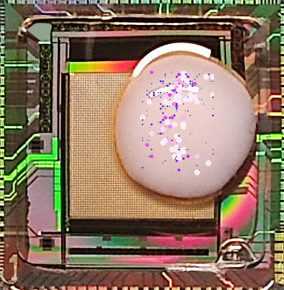
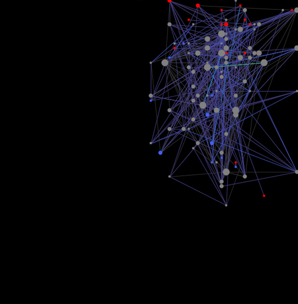
Hypercharge into new discoveries
-
– 3D chip captures 20-50x more signal than flat technologies
-
– Increase the robustness and sensitivity of your recording
-
– Discover hitherto unknown connectivity and network dynamics
-
– Allows for phenotypic screening and biomarker discovery
-
– Non-destructive to cytoarchitecture
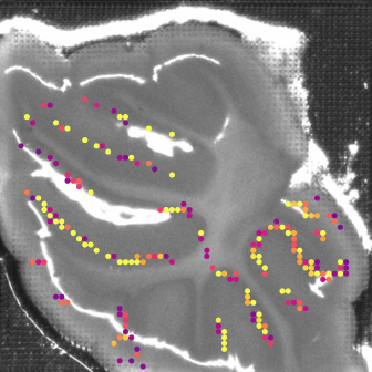
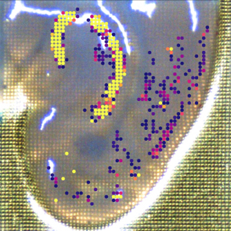
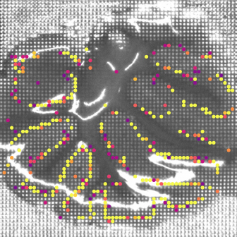
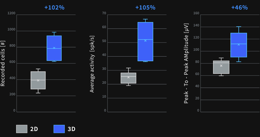
CorePlate™ 2D vs. 3D microchip on brain tissue slices
Brain slices 2D recording
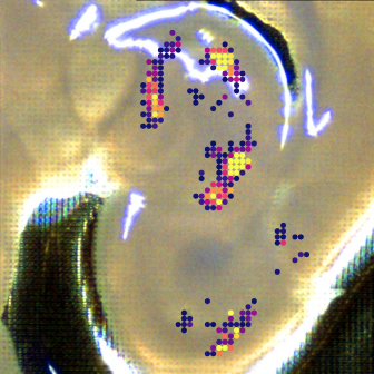
Brain slices 3D recording
Embrace the most nuanced inside view
-
– Record more cells and signals
-
– Bypass damaged cell surface layers of tissue slices
-
– Increase the robustness of your measurements
-
– Better reproducibility
On chip microfluidic channels for 3D model systems
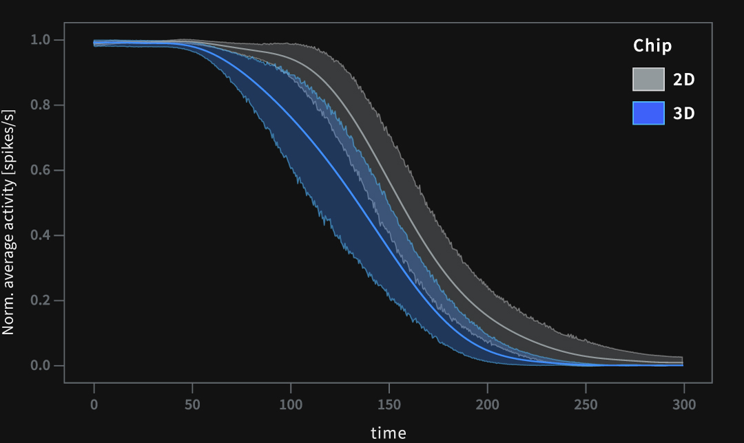
Response to sodium-channel blocker TTX
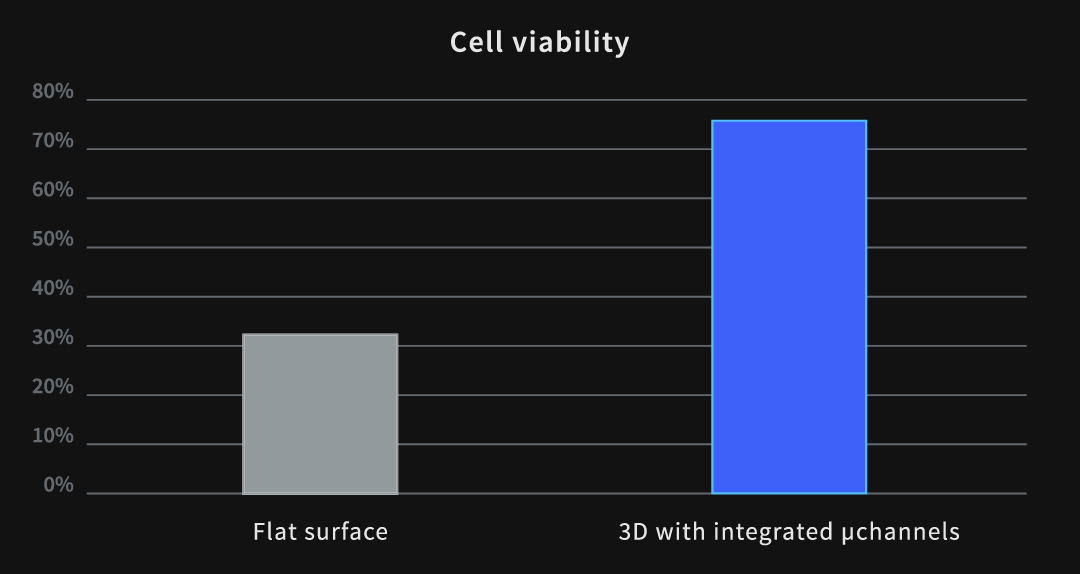
Developed for drug and phenotype screening
-
– Free nutrient and oxygen flow from below
-
– Increased cell viability and health
-
– Retains tissue integrity
-
– Efficient and easy compound delivery
Products
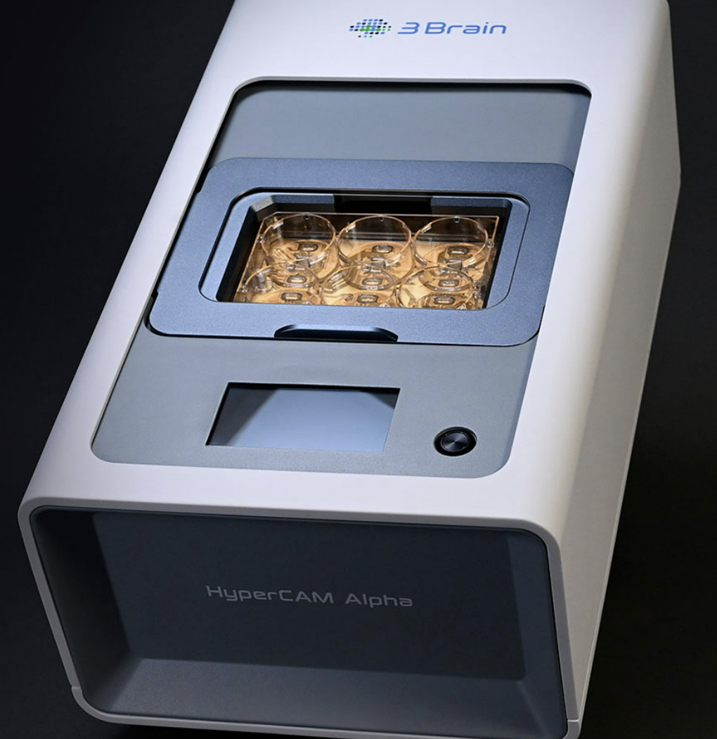
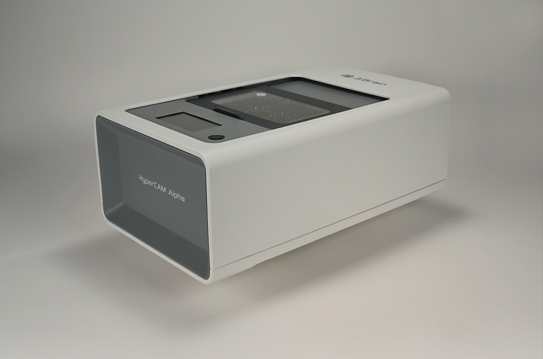
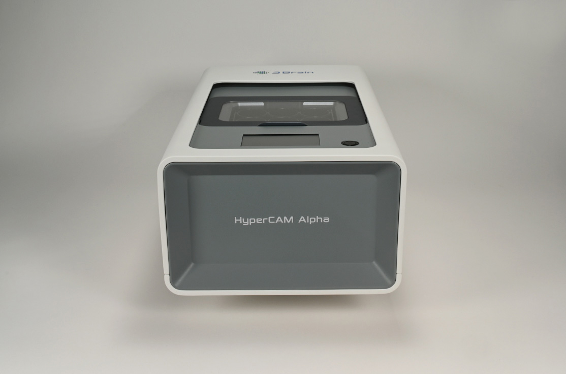
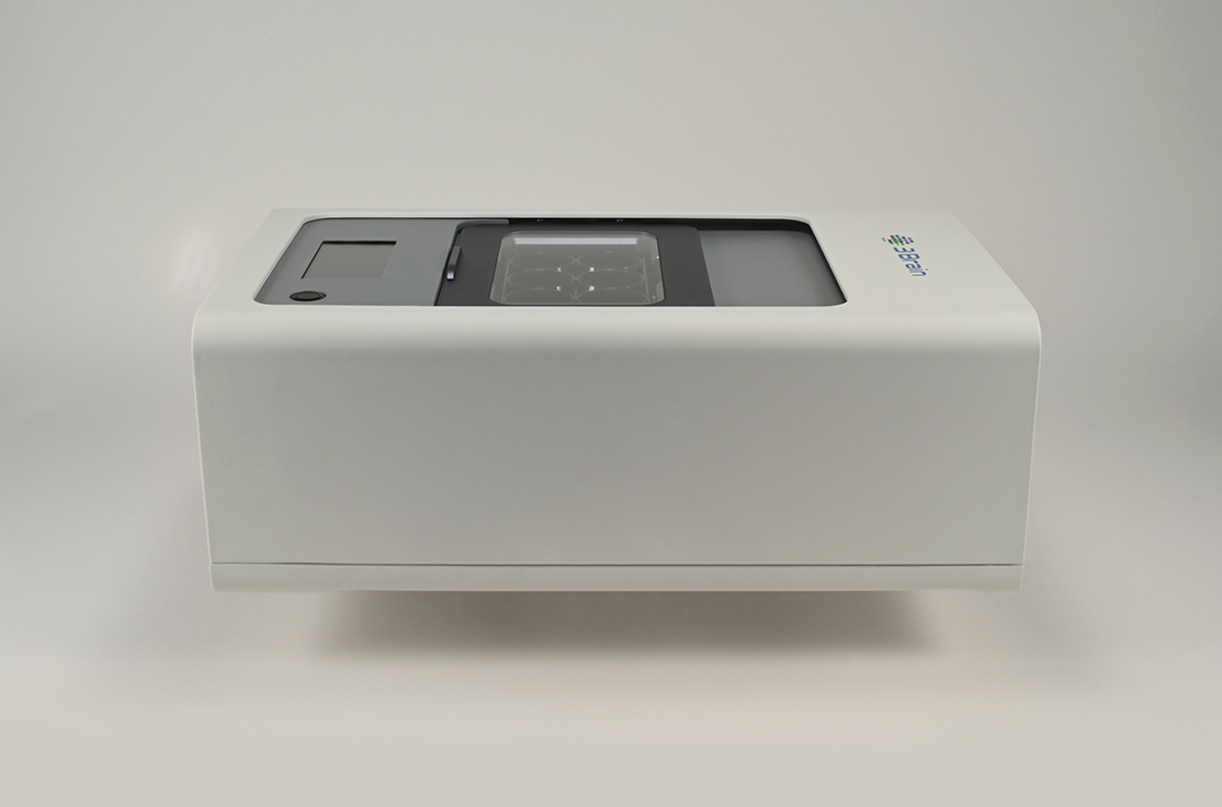
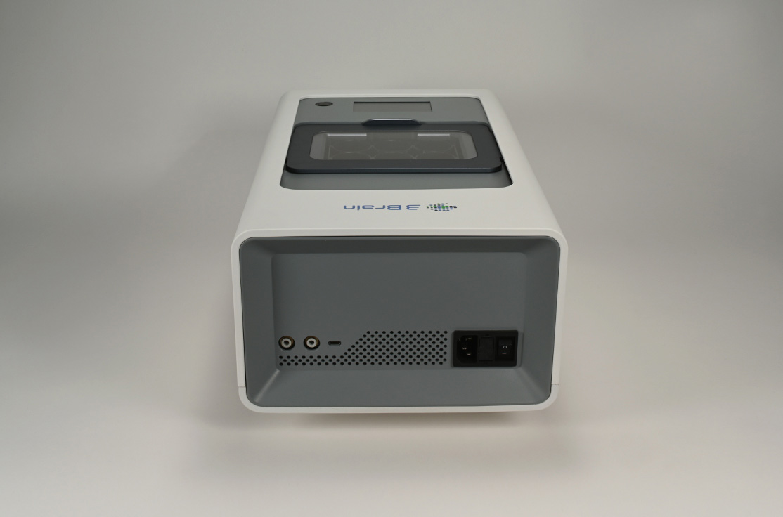
HyperCAM Alpha
HyperCAM Alpha multi-well system: A new frontier in functional imaging
The 6-well CorePlate™ -powered platform designed for both cell culture and tissue preparations
Overview
Your companion in functional cell screening
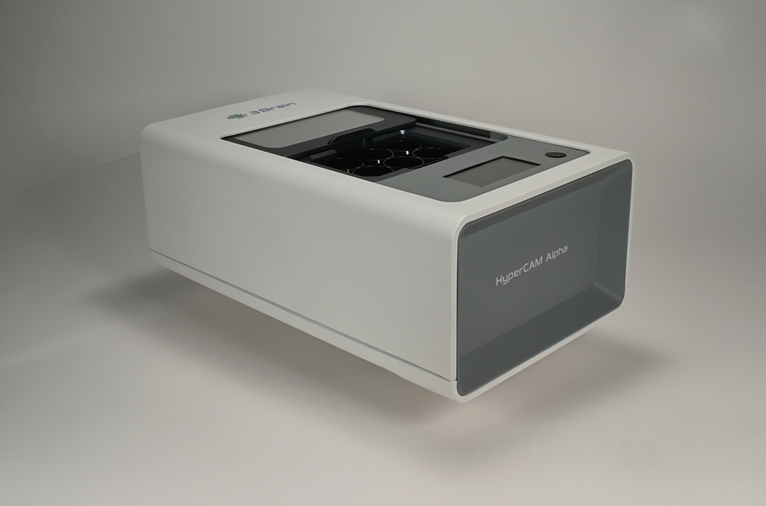
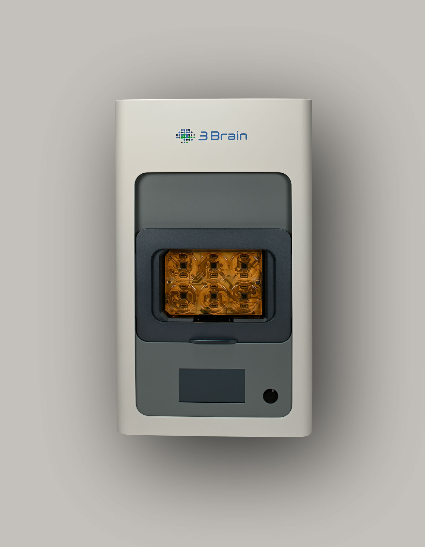
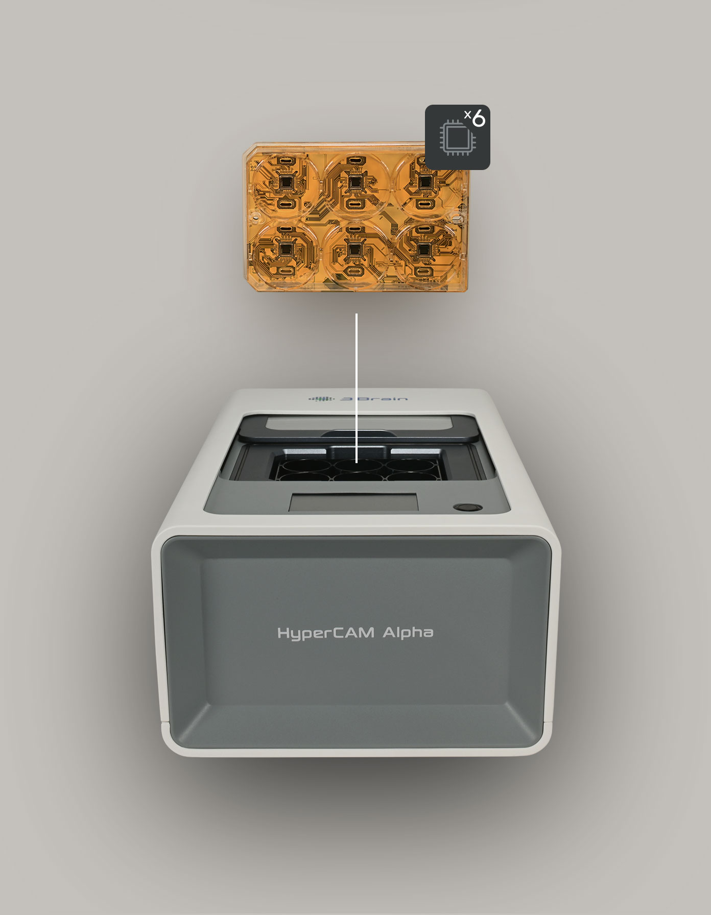
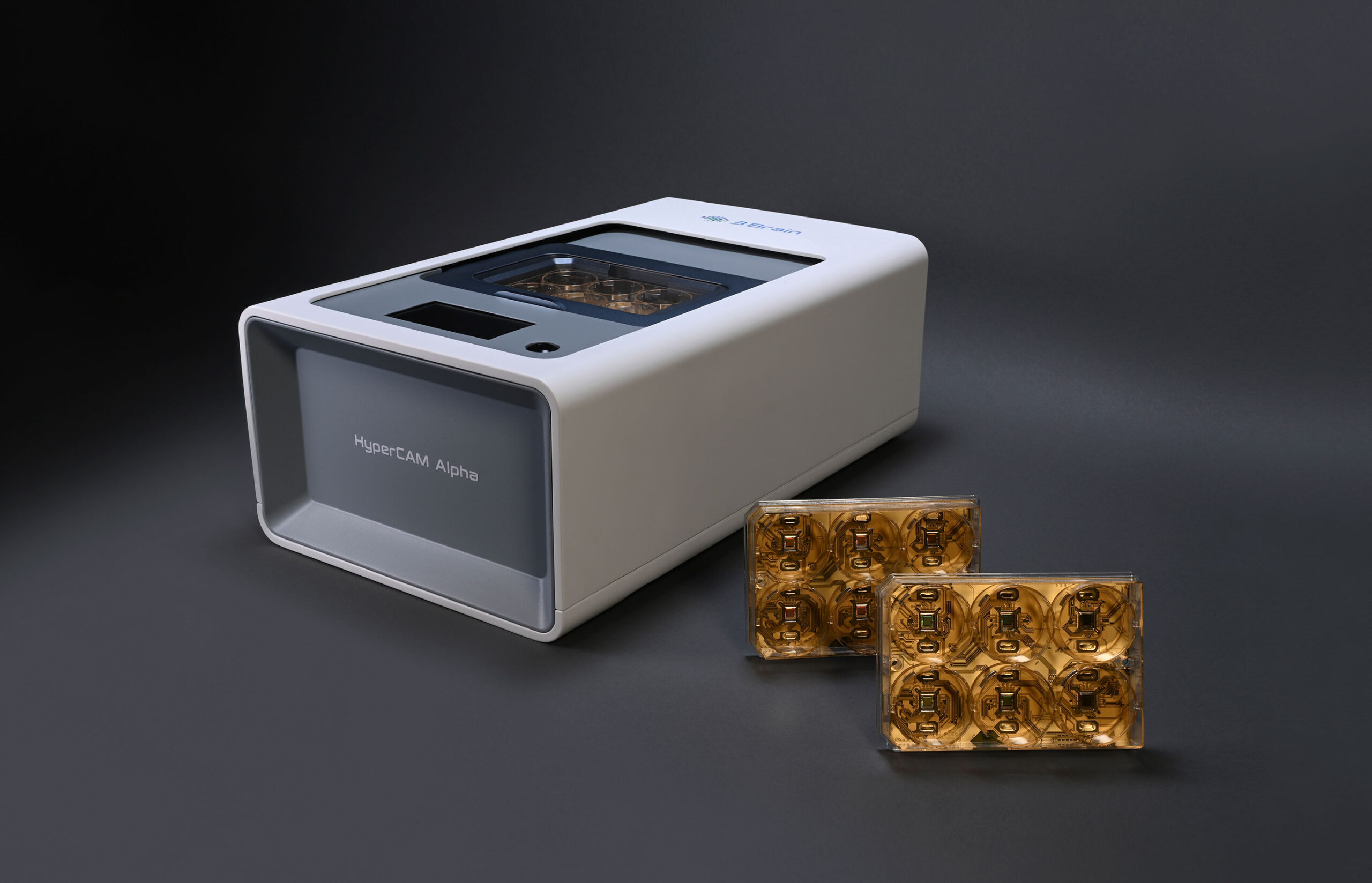
Thanks to the CorePlate™ technology, HyperCAM Alpha delivers high resolution functional screening platform in a multi-well plate format.
The controlled environment allows label-free kinetic in vitro assays. The large sensing area of the per-well integrated microchip provides room for measuring either cell cultures, brain slices or brain organoids.
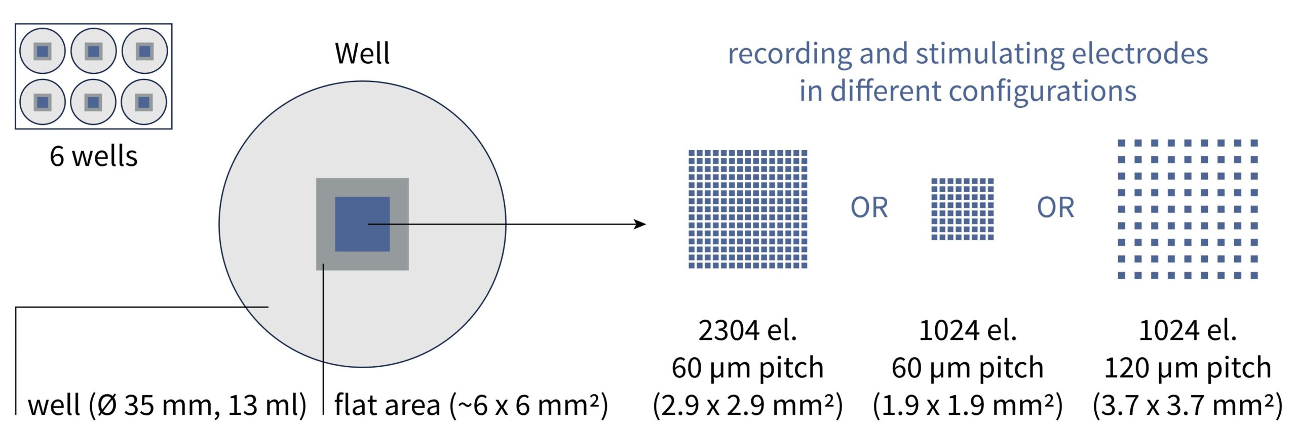
Alpha is equipped with the latest generation of FPGA accelerators to manage real-time high-resolution data from up to 13,824 (2,304 per well) simultaneously recorded electrodes.
Complex operations are handled by the sophisticated system so you can focus on measuring the functional properties of neurons.
Multiwell form factor with no compromise on data quality
Alpha accelerates your time-to-results without giving up any details
Parallelization does not have to be achieved at the cost of sacrificing data quality. Thanks to the CorePlate™ 6W 38/60, Alpha provides thousands of electrodes per-well that can be arranged in different recording configurations to satisfy any experimental needs. The high statistical quality of the data generated by each well reduces the overall needs of replicates. While also giving access to single-cell details with a spatio-temporal resolution comparable to that provided by our single well CorePlate™.
Smart and simple
A concentration of high-tech solutions to simplify experimental procedures
HyperCAM Alpha is designed to reduce experimental hassles and allows you to fully concentrate on the most important thing: your assay.
The CorePlate™ multiwell’s environment is regulated by accurately controlling the temperature, CO₂ saturation and humidity through the software or even the integrated touchscreen.
The incubation chamber is water-proof to protect instrument electronics from accidental spill-out or experimental misuse.
The combination with the latest BrainWave software allows to easily visualize and analyze data from CorePlate™ microchips.
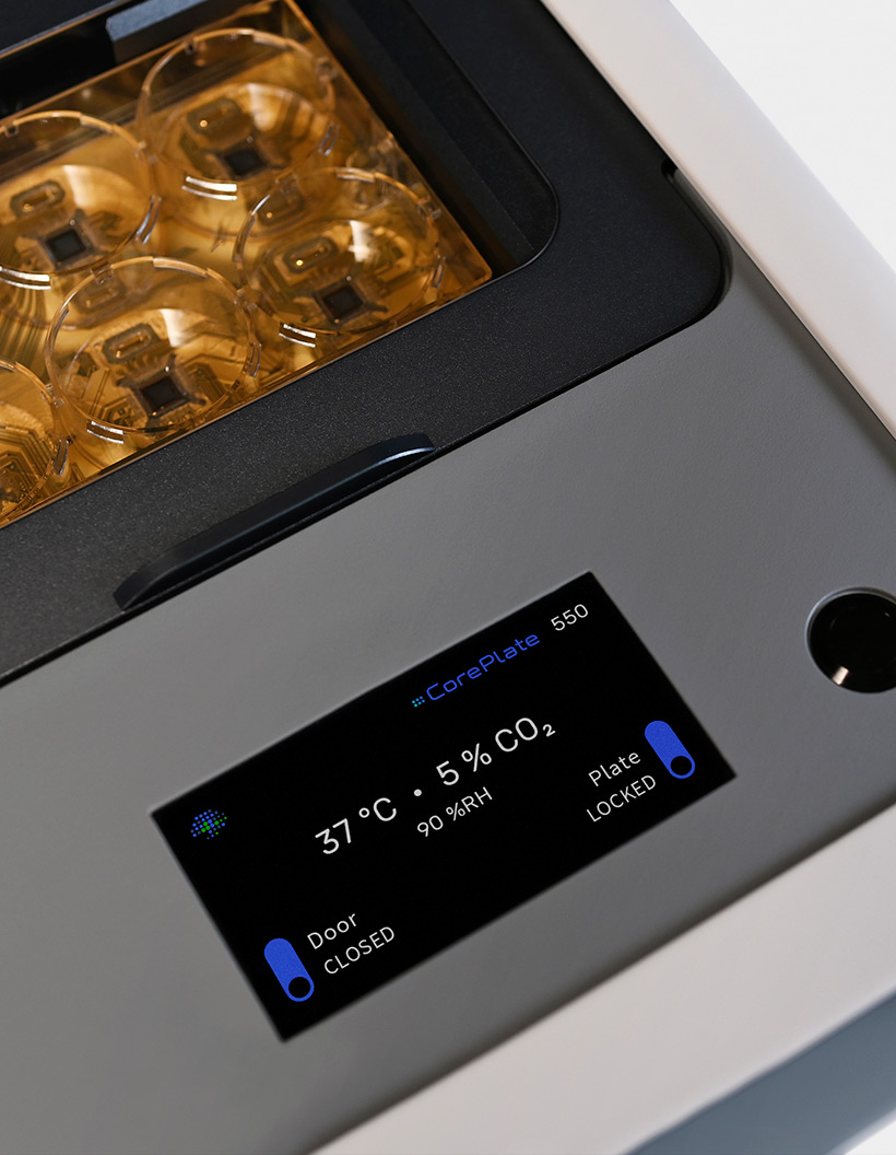
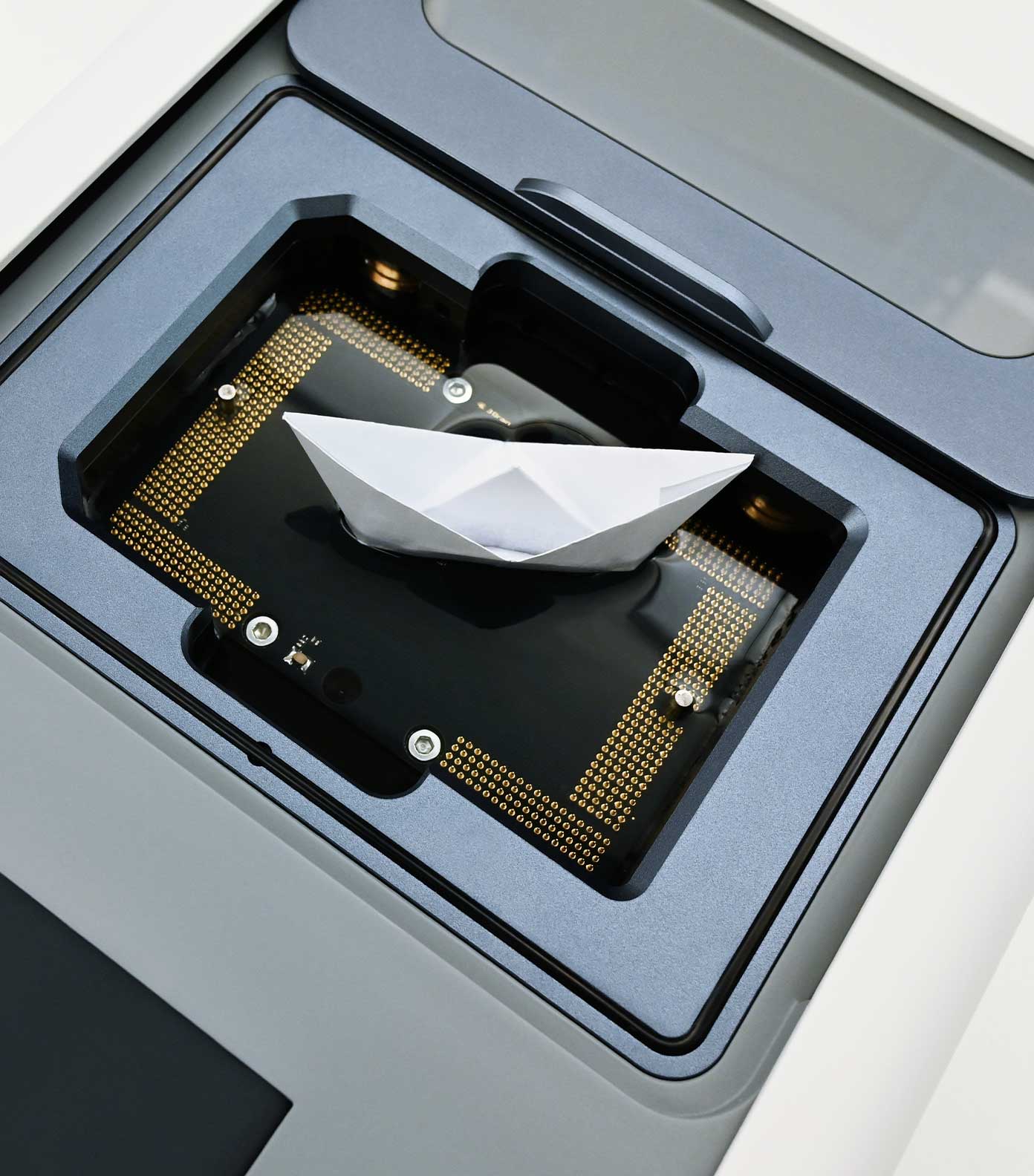
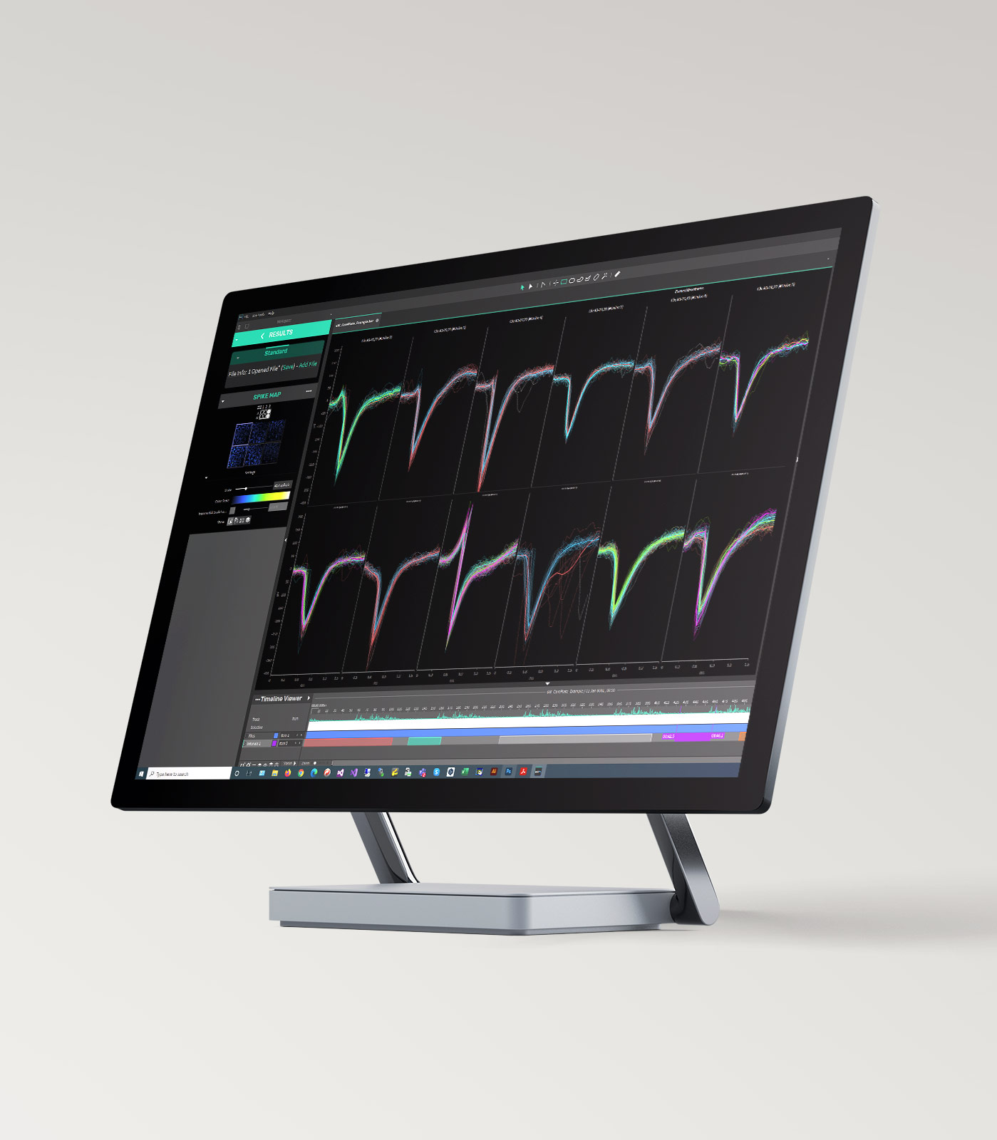
CorePlate™ Multiwell
Multiple cores for cell-based assays without compromises
After being the first introducing CMOS-based HD-MEA (in 2011) to overcome passive MEA limitations, we are now setting up the next standard by introducing the CorePlate™ technology, a multi-well plate HD-MEA with integrated processing power.

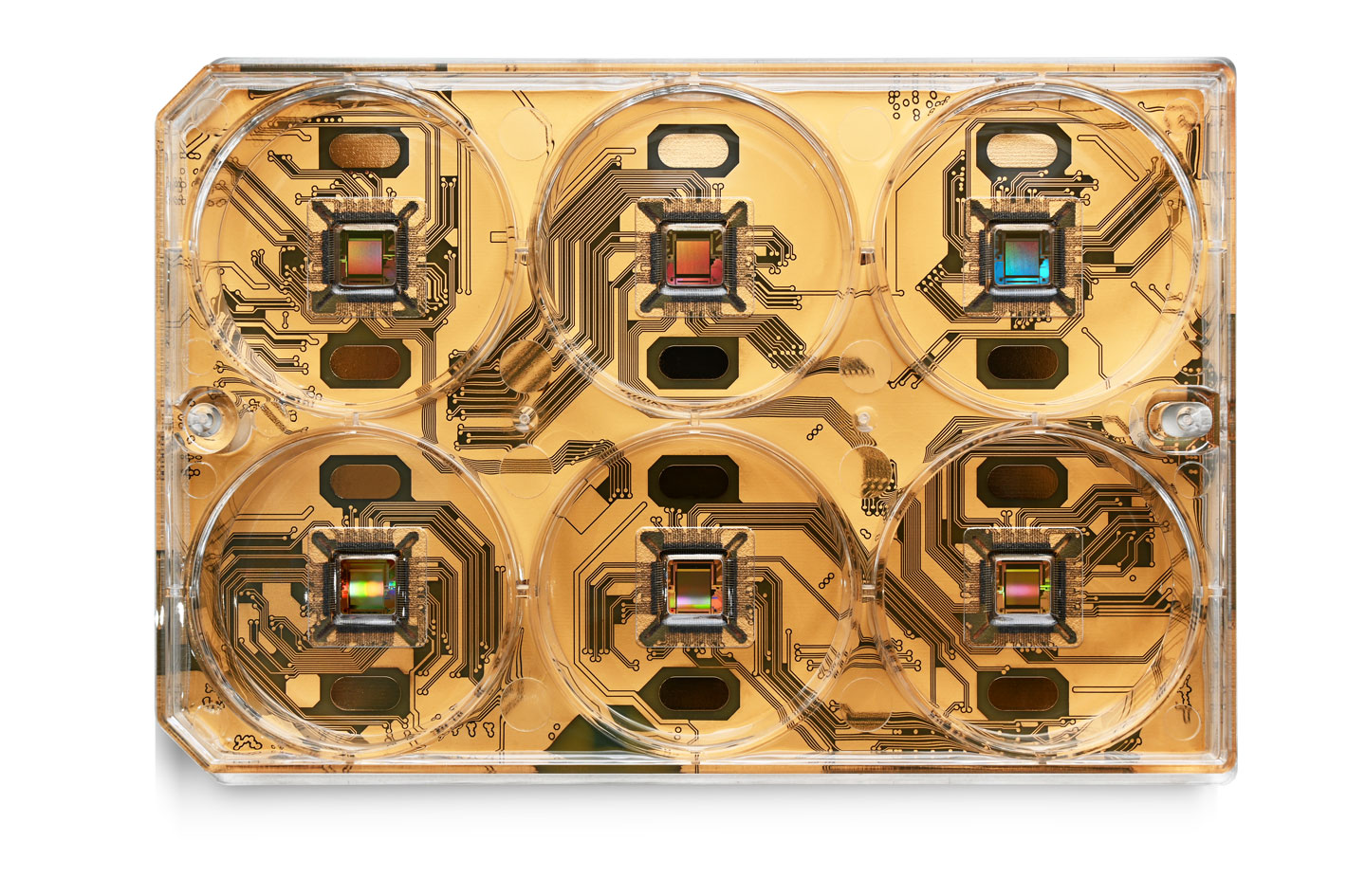
Flexible layout for both cell cultures and brain tissues
6-well CorePlate
The CorePlate™ powered 6-well integrates 4,096 bidirectional electrodes per well arranged in a 3.8 mm x 3.8 mm area. Up to 2,304 electrodes at 10 kHz or 1,024 electrodes at 20 kHz can be simultaneously recorded per well by selecting a subset of the 4,096-electrode array. Different recording subsets are available to either maximize the recorded area or the spatial resolution, and to suit both cell cultures and brain tissues.
Moreover, each of the 4,096 electrodes or combinations of those can be used to release an electrical stimulation to your sample.
Each microelectrode is 25 µm x 25 µm, with a 60 µm pitch. The internal diameter of the well reservoir is 35 mm, with a height of 13 mm.
BioCAM DupleX
Where the functional screening revolution begins
Introducing a fully bi-directional microelectrode array platform for 2D and 3D in vitro imaging
Electrophysiology in 3D
An ideal match: BioCAM DupleX and Khíron
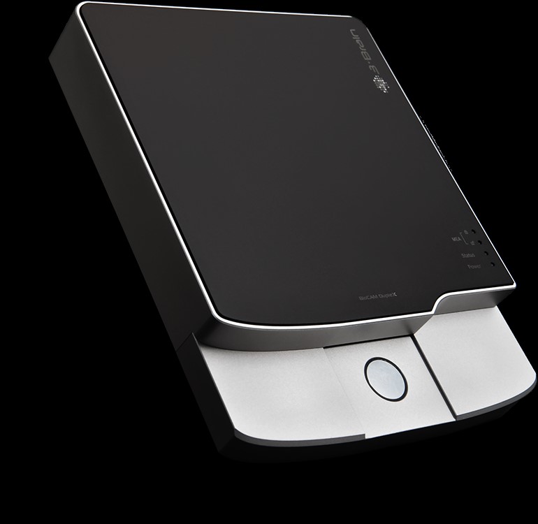
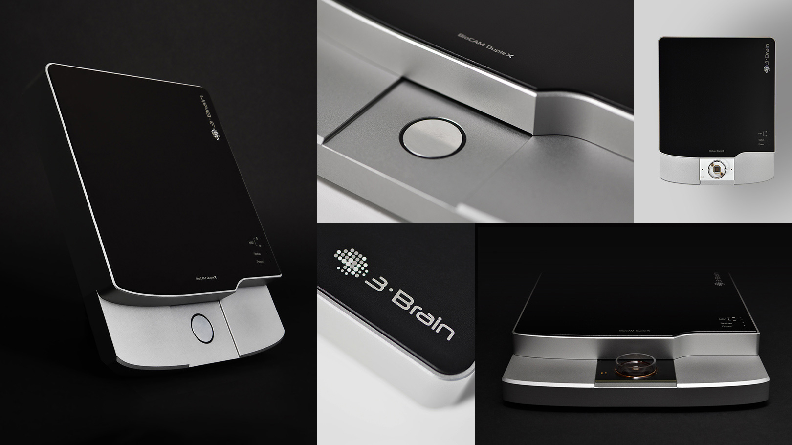
The BioCAM DupleX is the first Microelectrode Array platform of its kind able to power 3D high-density CMOS-MEA chips
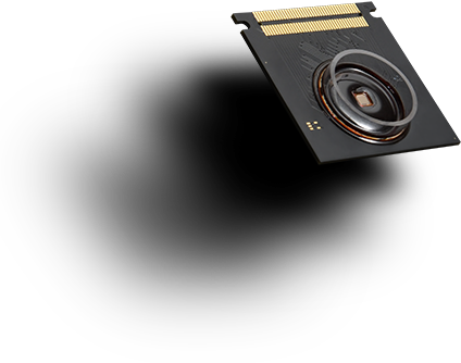
With CorePlate™ 1W 38/60, microchip family developed by 3Brain, the BioCAM DupleX can simultaneously record and stimulate from all 4,096 microelectrodes structured in a penetrating µNeedle grid.
These revolutionary µNeedle electrodes access information from inside all kinds of tissues – such as slices, organoids, spheroids and scaffolded 3D cultures – without damaging them, making the BioCAM DupleX the only platform for high-resolution in vitro 3D functional imaging.
Electrophysiology revolutionized
Complex electrophysiology setups, where the wiring of a single electrode to the amplifier can significantly change the noise level and impair the experiment, or where the interpretation of the signals coming from your setup is restricted to electrophysiological experts, are now a distant memory.
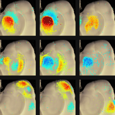
BioCAM DupleX makes setup as easy as possible by automating important settings and optimizing noise, thus making electrophysiology accessible to everyone. Complex electrophysiological traces are then converted into microscopy-like, easy-to-understand, fast-to-process functional images.
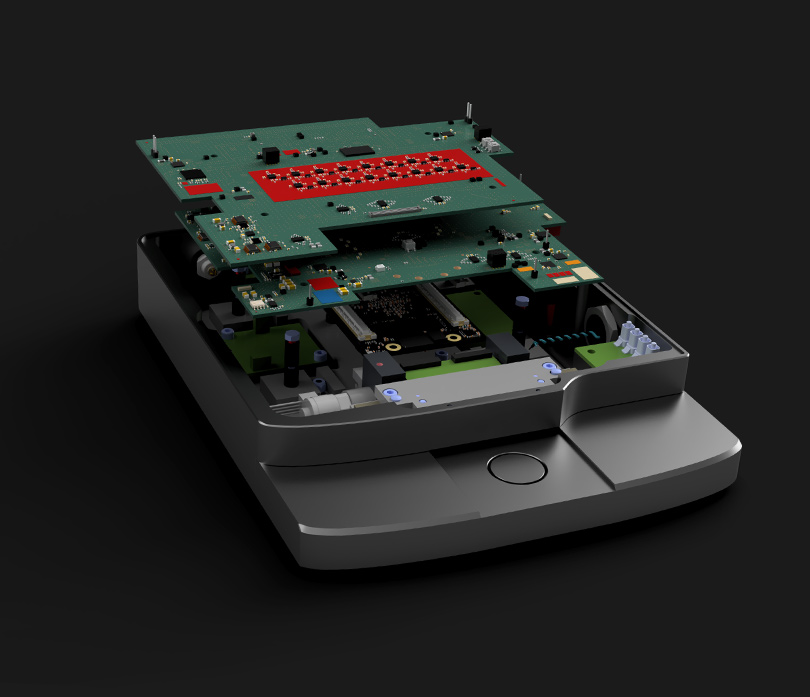
Sensing and actuation with 4,096 electrodes
Bidirectional platform
When paired with the HD-MEA from the Khíron family (CorePlate™1W 38/60), the BioCAM DupleX can use any of the recording sites to release an electrical stimulus, thus making it a fully bidirectional platform.
Coupled with BrainWave, our unique integrated software for recording, stimulation and real-time processing, BioCAM DupleX is the perfect solution for easily setting up complex experiments. Study functional connectivity, information processing or complex behaviors like learning and memory in neuronal networks – all without the need for external stimulating electrodes or third-party electronics.
Perfect fit in any setup
Compact design for maximum impact
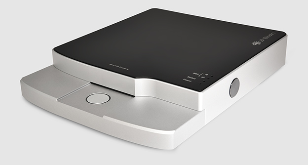
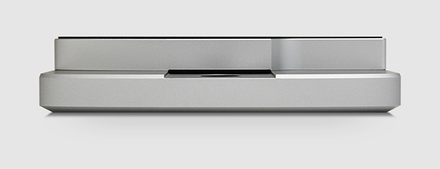
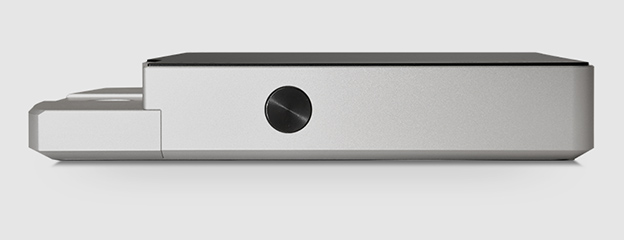
The BioCAM DupleX high-resolution microelectrode array (HD-MEA) platform is packed with features to enrich your research. At the same time, its small footprint makes for easy integration with instruments like microscope and perfusion or patch-clamp systems.
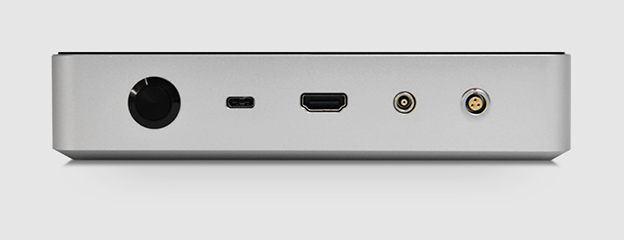
W 230 mm/9.5”
D 180 mm/7.08”
H 42 mm/1.65”
Single Well HD-MEAs
Single-Well HD-MEAs
High-density microelectrode arrays with large sensing areas
Our high-density microelectrode array (HD-MEA), also known as multielectrode array, features 4,096 low-noise microelectrodes that allow for simultaneous electrophysiological recording.
Overview
The perfect tool for your brain research
Investigating cellular networks requires time, expertise and effort to obtain the performance necessary to validate research hypotheses. With 3Brain as your technology partner, you can easily capture every cell signal in high resolution.
Our powerful CMOS MEA (microelectrode array / multielectrode array) system collects high-density electrophysiological data from preparations made of brain slices, neuronal tissue, human-derived stem cells,brain organoids, retinal tissue and cardiac cultures and tissues.
Different HD-MEA chip models for our microelectrode arrays are available to match your experimental needs in terms of resolution, field of view and stimulation capabilities.
Electrodes are coated with platinum to provide a stable and fully biocompatible surface. The quality of the recording is ensured by the outstanding signal-to-noise ratio, as you can see from our video samples gallery.
Recording and stimulating capabilities for all your applications
CorePlate™1W 38/60
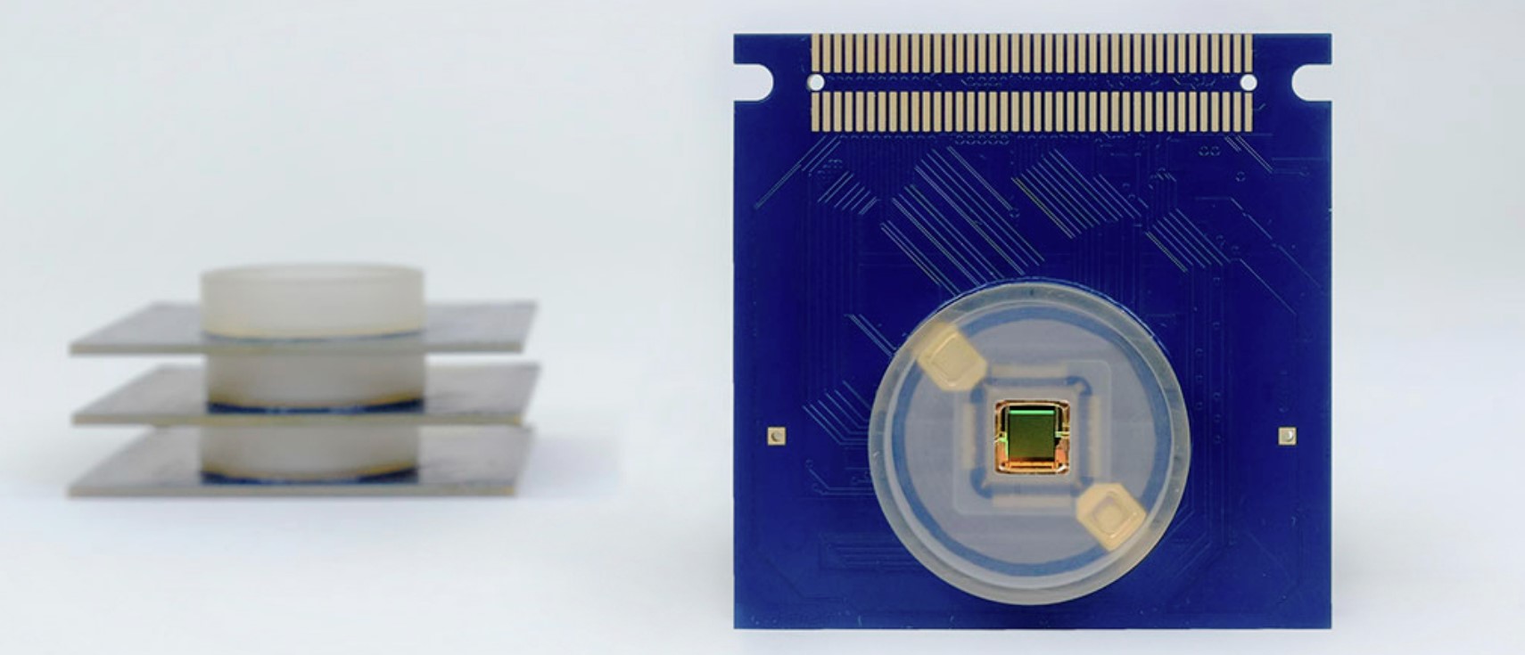
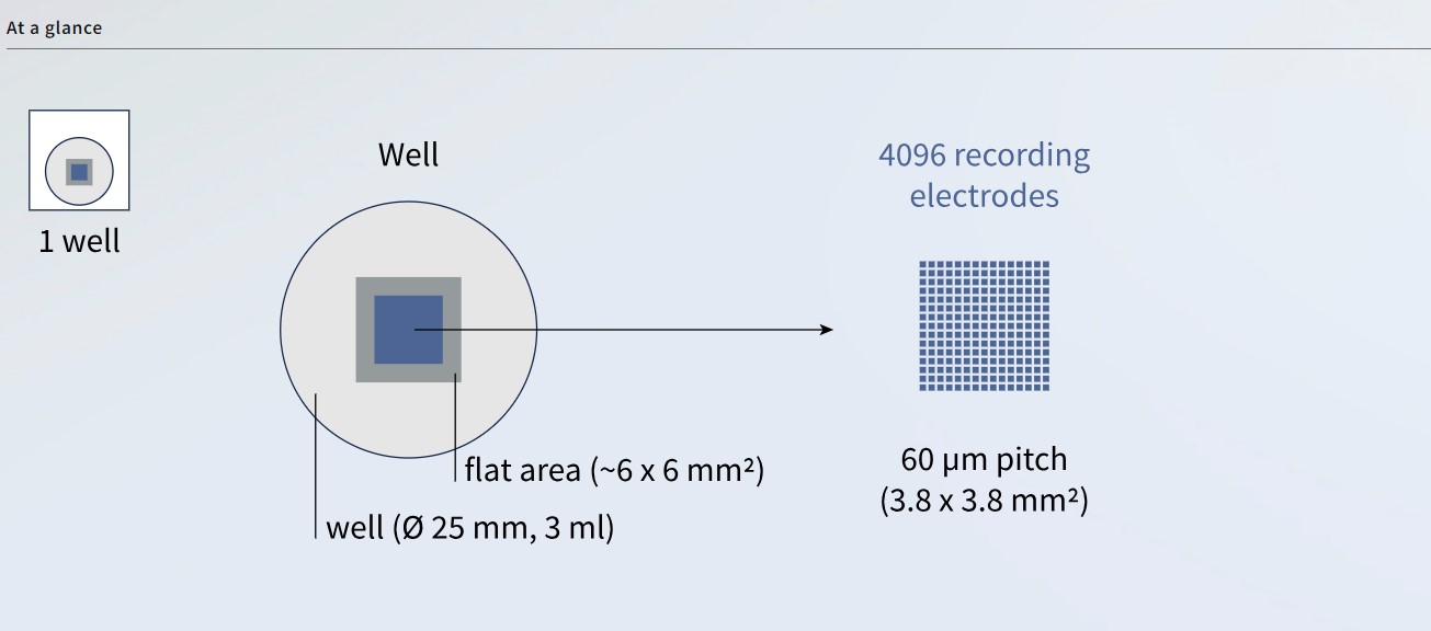
The CorePlate™1W 38/60 microelectrode array features 4,096 recording electrodes arranged in a 3.8 mm x 3.8 mm area, a perfect compromise for both cultures and brain tissues. Compared to previous chipset families, CorePlate™1W 38/60 achieves an improved signal-to-noise ratio by offering a recording mode where a subset electrode area can be accessed for ultra-low noise performance.
Moreover, every single recording electrode can also be used to release an electrical signal to your sample. The CorePlate™1W 38/60 provides a 3.8 mm x 3.8 mm grid of 4,096 electrodes for spatially accurate electrical stimulation at a cellular resolution.
Each microelectrode is 21 µm x 21 µm, with a 60 µm pitch. The internal diameter of the reservoir is 25 mm, with a height of 7 mm.
The CorePlate™1W 38/60 is based on the Khíron (Gen 3) chipset family and is compatible with BioCAM DupleX only.
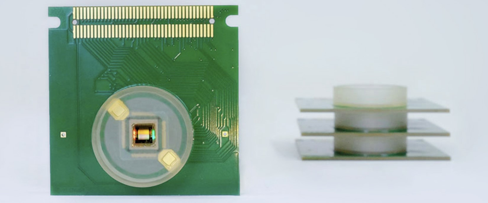
More space to accommodate large tissues
CorePlate™1W 27/42L
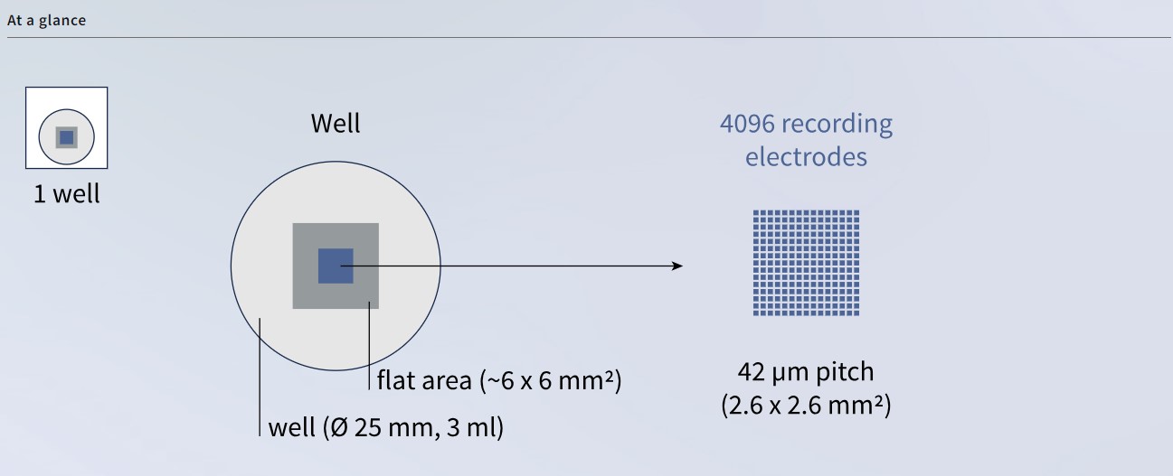
CorePlate™1W 27/42L has the same microelectrode recording array as the CorePlate™ 1W 27/42 (4,096 electrodes in a 2.67 mm x 2.67 mm area), but offers a larger surrounding flat area (~6 mm x 6 mm), which is ideal for positioning large tissues while guaranteeing optimal coupling with the microelectrode array.
Each microelectrode is 21 µm x 21 µm, with a pitch of 42 µm. The internal diameter of the reservoir is 25 mm, with a height of 7 mm.
The CorePlate™ 1W 27/42 is based on the Artemis (Gen 2) chipset family and can be used with BioCAM X and BioCAM DupleX.
Largest sensing area and stimulation capabilities
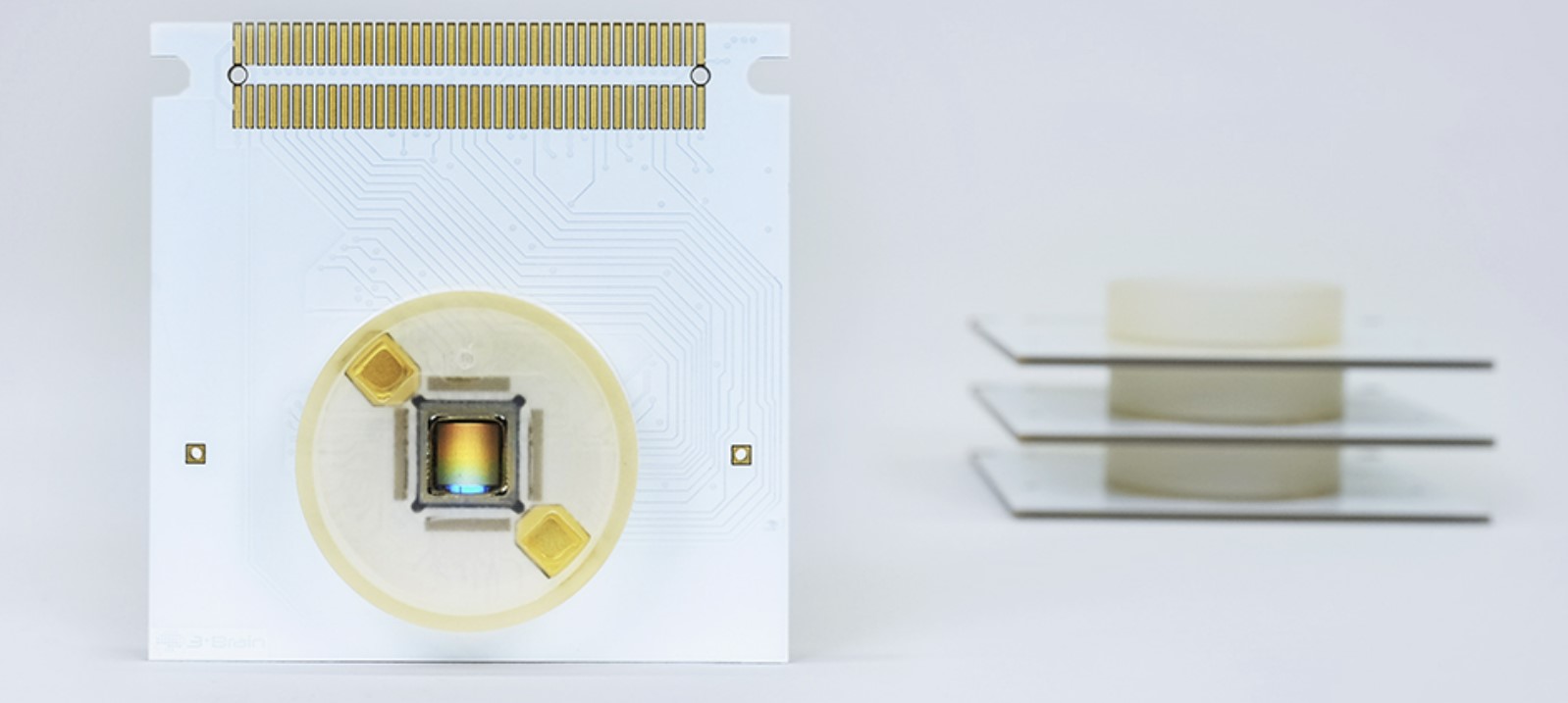
CorePlate™ 1W 51/81
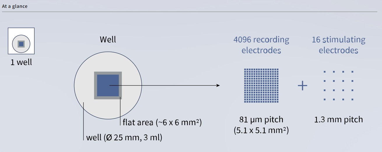
The CorePlate™ 1W 51/81 features a large sensing area (5.12 mm x 5.12 mm) with an increased electrode pitch of 81 µm. This makes it possible to record electrophysiological activity from large portions of neuronal tissue while maintaining a high spatial resolution.
The CorePlate™ 1W 51/81 model also features a secondary array for stimulation with 16 microelectrodes, arranged in a 4 x 4 uniform grid, that are interspersed between the recording electrodes.
The electrode size for both recording and stimulating electrodes is 21 µm x 21 µm. The internal diameter of the reservoir is 25 mm, with a height of 7 mm.
CorePlate™ 1W 51/81 is based on the Artemis (Gen 2) chipset family. It can be used to its full extent with BioCAM X. Whereas when it is plugged into the BioCAM DupleX, its stimulation array is not supported and only recording is possible.
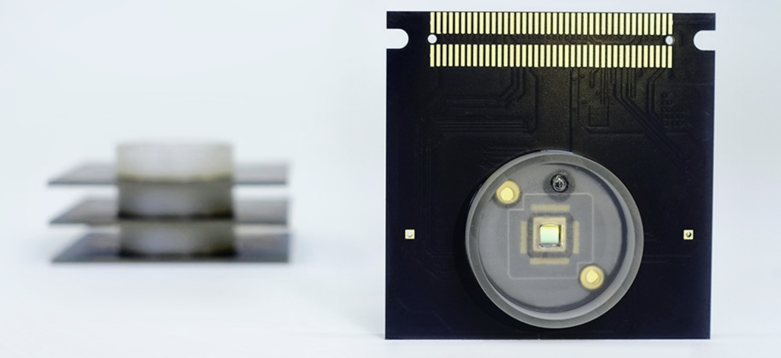
Ideal for cell cultures
CorePlate™ 1W 27/42
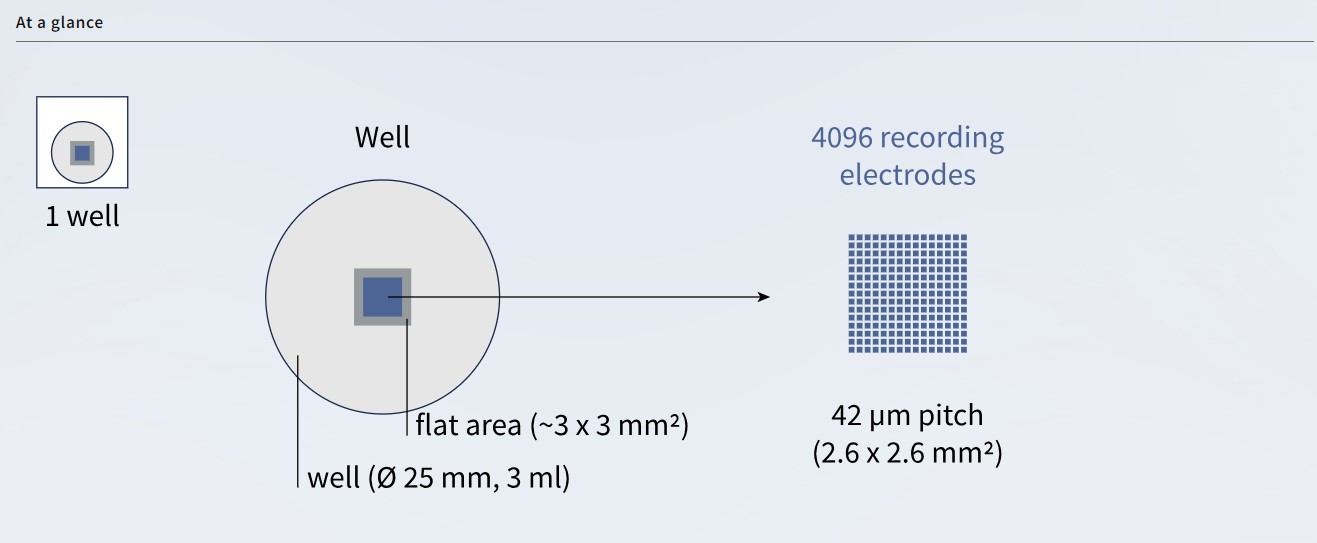
CorePlate™ 1W 27/42 is a high-density microelectrode array (HD-MEA) integrating 4,096 recording electrodes on a small and compact area of 2.67 mm x 2.67 mm, centered on a flat area of ~3 mm x 3 mm. This arrangement is ideal in reducing the number of seeded cells.
Each microelectrode is 21 µm x 21 µm, with a pitch of 42 µm. The internal diameter of the reservoir is 25 mm, with a height of 7 mm.
The CorePlate™ 1W 27/42 is based on the Apollo (Gen 1) chipset family and can be used with BioCAM X and BioCAM DupleX.
Application
Neuronal cell culture and stem cell cultures
Explore new pathways in brain research and drug discovery
Models
– Human iPSC-derived neuronal culture – Neural progenitor derived cultures – Primary neuronal cell culture
Brain Organoids and 3D Neuronal Cultures
Generate electrophysiology maps for precise brain imaging in vitro. Record electrophysiological activity in brain spheroids, brain organoids and retinal organoids.
Models
– Brain/CorticalOrganoids – Retinal Organoids – Spheroids – Scaffold/matrix-based 3D cultures
Brain Slice Electrophysiology
Boost neuronal circuitry studies through functional brain tissue imaging
Models
– Acute brain slices – Organotypic brain slices
Ex-vivo acute retinal explant
Shed light on retinal activity in vitro. Record responses to light stimuli and generate retinal ganglion cells (RGCs) mosaics.
Models
– Whole mount retina explants – Primate and non-primate retina tissues
Cardiomyocyte Cells
Perform in vitro electrocardiograms to gain functional and spatiotemporal insights from cardiac cells. Record electrophysiological activity in cardiomyocytes cultures, cardiac spheroids, and cardiac organoids.
Models
– Primary cardiomyocytes culture – Human induced pluripotent stem-cell derived cardiomyocytes – Explanted heart tissue
Pharma on Chip
Revolutionizing in vitro preclinical screening
Models

