Cellames Cell Analysis Team
Abstract
Analysis of Field Potential Parameters in iCell Cardiomyocytes Using the CFPS-32 System
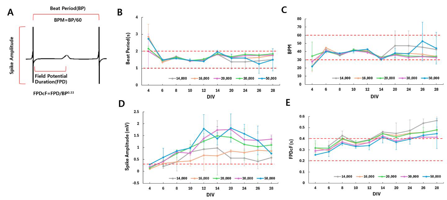
A total of 14,000–50,000 cardiomyocytes per well were cultured on CITO-16W01E-SGL cell chip up to DIV28, and field potentials were measured using the CFPS-32 system. (A) The key field potential parameters of cardiomyocytes were analyzed using the CFPS-32 software. (B) Cardiomyocyte beat period stabilized between 1-2 seconds starting from DIV6. (C) A consistent BPM around 30-60 was observed across various densities. (D) While the spike amplitude generally showed high variability, it tended to increase with higher cell densities, and after DIV8, it remained above 0.3 mV. (E) FPDcf stabilized around DIV10 and tended to increase with longer incubation periods. All parameters were the most stable between DIV10-12. The red dotted lines indicate the acceptable range for each parameter.
Keyword
iCell Cardiomyocytes, CFPS-32, Cardiotoxicity, Nifedipine
Main Text
Workflow for Field potential measurement using CFPS-32

iCell Cardiomyocytes on CITO-16W01E-SGL Cell Chip
iCell cardiomyocytes of DIV7 by seeding 20,000 cells on the recording electrode
The recording electrode of the CITO-16W01E-SGL cell chip is coated with fibronectin and then incubated with iCell cardiomyocytes. During incubation, the morphology and beating of the iCell cardiomyocytes can be easily observed using transparent electrodes.
Field Potential Measurement of iCell Cardiomyocytes

Analysis of Field Potential Parameters in iCell Cardiomyocytes Using the CFPS-32™ System
A total of 14,000–50,000 cardiomyocytes per well were cultured on CITO-16W01E-SGL cell chip up to DIV28, and field potentials were measured using the CFPS-32TM system. (A) The key field potential parameters of cardiomyocytes were analyzed using the CFPS-32 software. (B) Cardiomyocyte beat period stabilized between 1-2 seconds starting from DIV6. (C) A consistent BPM around 30-60 was observed across various densities. (D) While the spike amplitude generally showed high variability, it tended to increase with higher cell densities, and after DIV8, it remained above 0.3 mV. (E) FPDcf stabilized around DIV10 and tended to increase with longer incubation periods. All parameters were the most stable between DIV10-12. The red dotted lines indicate the acceptable range for each parameter.
Analysis of Cardiotoxicity after Drug Treatment

Analysis of Field Potential Changes in iCell Cardiomyocytes with Nifedipine (Ca+ channels blocker)
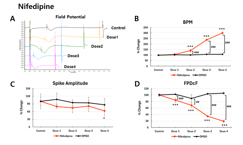
A total of 40,000 cardiomyocytes per well were cultured on CITO-16W01E-SGL cell chip until DIV12, and field potentials before and after nifedipine treatment were compared and analyzed. (A) The field potential waveform showed a decreased beat period with increasing dose of nifedipine. (B) The PBM significantly increased as the nifedipine dose increased. (C) The spike amplitude remained unchanged following nifedipine treatment. (D) FPDcF was significantly shortened with higher dose of nifedipine. Statistical analyses were performed using the Dunnett's test (***p<0.001, ###p<0.001).

Analysis of Field Potential Changes in iCell Cardiomyocytes with E-4031 (K+ channels blocker)
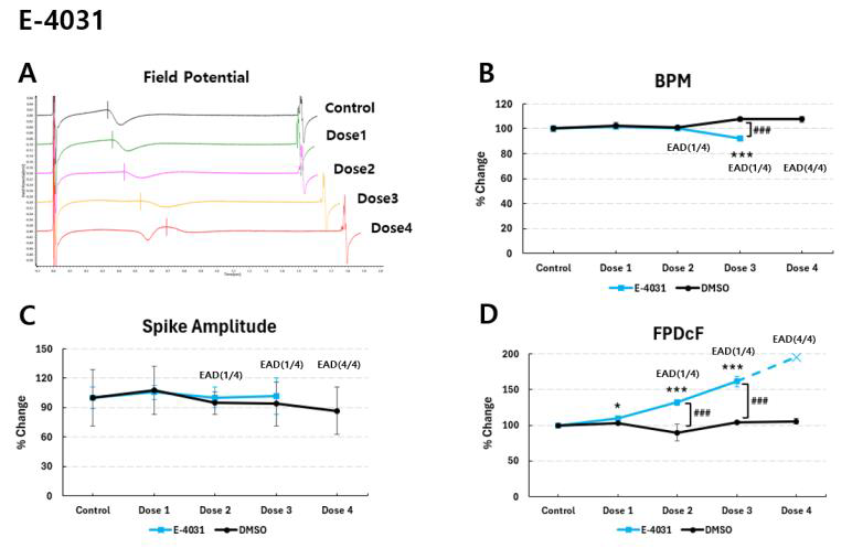
A total of 40,000 cardiomyocytes per well were cultured on CITO-16W01E-SGL cell chip until DIV12, and field potentials before and after E-4031 treatment were compared and analyzed. (A) The field potential waveform showed an increased beat period with increasing dose of E-4031. (B) The PBM significantly decreased as the E-4031 dose increased. (C) The spike amplitude remained unchanged following E-4031 treatment. (D) FPDcF was significantly prolonged with higher doses of E-4031. A key observation was the occurrence of EADs (Early Afterdepolarizations) at higher doses, with EADs observed in all wells (4/4) at Dose 4. CFPS-32 system demonstrated the ability to detect drug-induced proarrhythmic events in cardiomyocytes. Statistical analyses were performed using the Dunnett's test (***p<0.001, ###p<0.001).
Bibliographic Information
Research Ethics Related Information
Licenses
Attribution (CC BY)
