
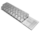
CIMS-Plate™ CELLAMES Electrode Cell Chip
Features- Each of the 16 channels has a single transparent circular sensing area with a radius of 250μm patterned on the slide glass (25 mm x 100 mm).
- The transparent circular sensing electrodes can be separated into 1, 2, 4, or 8 wells of polystyrene chamber which is removable from the electrode patterned glass substrate.
- The removable chamber is convenient for staining and microscopic examination without cell transfer.
- Monitoring of Cell Adhesion, Migration, Spreading
- Cellular Proliferation, Growth, or Differentiation
- Apoptosis, Necrosis
- Cytotoxic, Cytopathic effect
- Correlation with Microscopy
- CIMS-32
- CIMFS-32
| Model Name |
Electrode Material |
Electrode Transparency |
Growth Area / Well(cm2) |
Working Volume / Well(mL) |
Number of Well |
Electrode Type |
|---|---|---|---|---|---|---|
| CITO- 16W01E-IFE |
ITO(Indium Tin Oxide) |
Transparent | 0.33 | 0.2 ~ 0.3 | 16 | IFE(Interdigitated and Focused-shaped Electrode) |
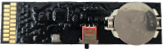
| CPCB -16W01E-IFE |
Alloys | Opaque | 0.33 | 0.2 ~ 0.3 | 16 | IFE |
| Model Name |
Type | Simulator | Usage |
|---|---|---|---|
| CMDL-16WQC-IMP | Model Cell | Impedance for CIMS-32/CIMFS-32 | QC and Calibration |
CITO-16W01E-IFE
CPCB-16W01E-IFE
CMDL-16WQC-IMP
Electrode Shape
IFE Electrode
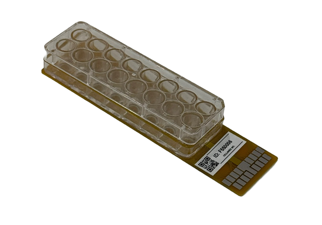
CFPS-Plate™ CELLAMES Electrode Cell Chip
Features- Each of the 16 channels has a single transparent circular recording electrode with a radius of 250 μm and including reference electrode patterned on the slide glass (26 mm x 100 mm).
- The transparent circular sensing electrodes can be separated into 1, 2, 4, or 16 wells of polystyrene chamber
- The electrode patterned on ITO(Indium Tin Oxide) glass substrate.
- Convenient for optical microscope examination (Transparent ITO electrode materials)
- Non-destructive electrical analysis of cardiomyocyte cells
- Field potential measurement
- Cardiomyocyte cells cytotoxicity test
- Supports organoid analysis with integrated MEA cell chips
- CFPS-32
- CIMFS-32
| Model Name |
Electrode Material |
Electrode Transparency |
Growth Area / Well(cm2) |
Working Volume / Well(mL) |
Number of Well |
Electrode Type |
|---|---|---|---|---|---|---|
| CITO- 08W01E-SGL |
ITO(Indium Tin Oxide) |
Transparent | 0.98 | 0.2 ~ 0.6 | 8 | Circular Disk Type (1 recording, 1 reference) |
| CITO- 16W01E-SGL |
ITO | Transparent | 0.33 | 0.2 ~ 0.3 | 16 | Circular Disk Type |
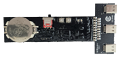
| CPCB- 16W01E-SGL (Coming Soon) |
Alloys | Opaque | 0.33 | 0.2 ~ 0.3 | 16 | Circular Disk Type |
| CITO- 02W16E-10A |
ITO | Transparent | 2.54 | 0.2 ~ 2 | 2 | MEA(100μm) |
| Model Name |
Type | Simulator | Signal Type | Usage |
|---|---|---|---|---|
| CMDL-01WQC-MEA | Model Cell | LFP for CFPS-32/CIMFS-32 | Real Signal of Cardiomyocyte | QC and Calibration |
| CMDL-01WQC-NR | Model Cell | LFP for CFPS-32-NR | Real Signal of Neuron Cell | QC and Calibration |
CITO-08W01E-SGL
CITO-16W01E-SGL
CPCB-16W01E-SGL
CITO-02W16E-10A
CMDL-01WQC-MEA
CMDL-01WQC-NR
Electrode Shape

SGL(Circular Single Electrode)

MEA
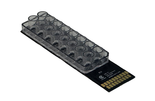
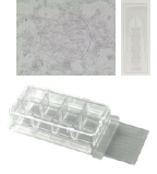
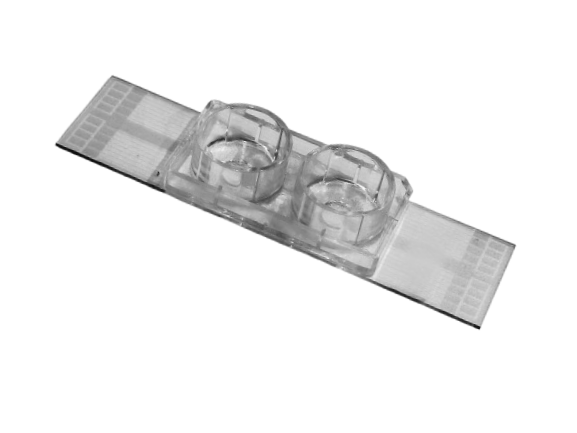
CFPS-Plate™ (2-Well Type for 3D-Structrued Cell Analysis)
Features- Each of the 2 channels has 16 circular recording electrodes with a diameter of 100 μm, along with a reference electrode patterned on the slide glass (26 mm × 100 mm).
- Larger well size optimized for effective 3D cell analysis.
- The electrode patterned on ITO(Indium Tin Oxide) glass substrate.
- The holder and silicone net help keep the cells in place, ensuring contact with the electrodes.
- Convenient for optical microscope examination (Transparent ITO electrode materials)
- Non-destructive electrical analysis of 3D cells
- Field potential measurement
- Cell cytotoxicity test
- Supports organoid analysis with MEA cell chips
- CFPS-32
- CIMFS-32
| Model Name |
Electrode Material |
Electrode Transparency |
Growth Area / Well(cm2) |
Working Volume / Well(mL) |
Number of Well |
Electrode Type |
|---|---|---|---|---|---|---|
| CITO- 02W16E-10A |
ITO | Transparent | 2.54 | 0.2 ~ 2 | 2 | MEA(100μm) |
| CITO- 02W16E-06A (Coming Soon) |
ITO | Transparent | 2.54 | 0.2 ~ 2 | 2 | MEA(60μm) |
CITO-02W16E-10A
CITO-02W16E-06A
Silicone Net & Holder
- Cells are kept well-attached to electrodes without detachment using Silicone net and Holder.
- Enhanced transparency for improved microscopic observation.
- Silicone nets are provided in various height options: (0.2mm, 0.5 mm, 1 mm, 1.5 mm, 2 mm)
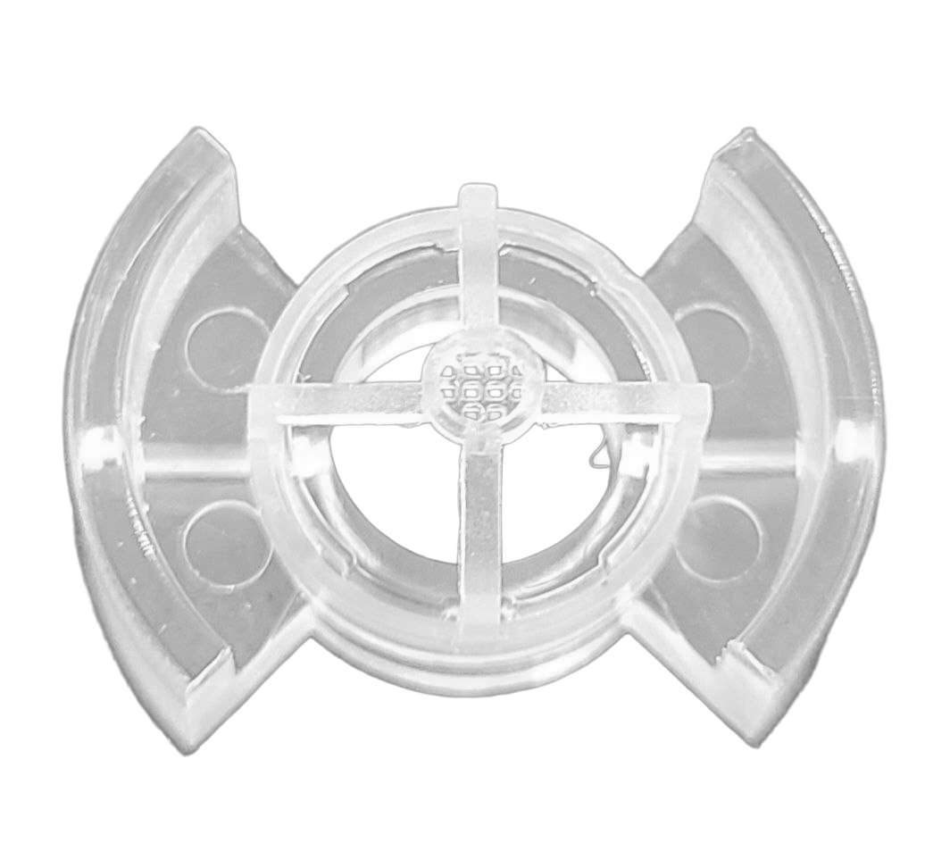
Silicone Net and Holder
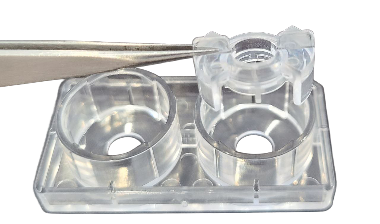
Assembly with cultureware